
Precision Treatment for Coronary Artery Disease and Complex Heart Blockages
All treatment decisions are guided by a physiology-first, imaging-confirmed strategy aligned with contemporary international interventional cardiology standards.
What Is Coronary Artery Disease?
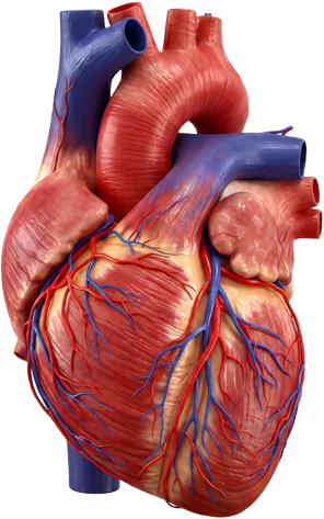
Coronary artery disease is one of the most common cardiovascular conditions worldwide. It occurs when the arteries supplying blood to the heart become narrowed or blocked due to plaque build-up, reducing blood flow to the heart muscle and leading to chest pain, breathlessness, fatigue, or, in severe cases, a heart attack.
The severity of symptoms does not always match the severity of blockage, which is why modern cardiology has moved beyond simple visual assessment. Modern treatment decisions are based on a combination of anatomical imaging, physiological assessment, and patient-specific risk profiling to ensure every patient receives the most appropriate and evidence-based treatment strategy.
Other Services
Conditions Treated
Coronary Artery Disease (CAD)
Plaque build-up inside the coronary arteries reducing blood flow to the heart and increasing the risk of angina and heart attack.
Stable Angina
Chest discomfort during exertion due to reduced blood supply to the heart muscle.
Unstable Angina
Chest pain occurring at rest or becoming progressively worse, requiring urgent evaluation.
Acute Coronary Syndrome
A spectrum of conditions including heart attack and unstable angina caused by sudden reduction in blood flow to the heart.
Stent Failure or Restenosis
Re-narrowing of a previously treated artery requiring reassessment and advanced re-intervention strategies.
Heart Attack (Myocardial Infarction)
A life-threatening event caused by complete blockage of a coronary artery requiring immediate intervention.
Multivessel Coronary Disease
Significant narrowing in multiple coronary arteries requiring complex decision-making for optimal treatment.
Chronic Total Occlusion (CTO)
A complete long-standing blockage of a coronary artery that requires specialised advanced techniques to treat.
Calcified Coronary Disease
Severe hardening of coronary arteries due to calcium build-up, making standard stenting techniques more challenging.
Precision Diagnosis Before Intervention
Coronary Angiography
A key diagnostic procedure using X-ray imaging to visualise the coronary arteries and identify blockages. It remains the foundation of coronary assessment but is always interpreted alongside functional and imaging data.
Intracoronary Physiology (FFR and iFR)
Physiological testing measures the actual impact of coronary narrowing on blood flow. This ensures only blockages causing true limitation of blood supply are treated, avoiding unnecessary procedures.
Intravascular Imaging (IVUS and OCT)
High-resolution imaging performed from inside the artery, allowing precise assessment of plaque composition, vessel size, and stent optimisation. Essential in complex or calcified disease.
Coronary Interventions
Percutaneous Coronary Intervention (PCI)
A minimally invasive catheter-based procedure using balloons and stents to open blocked or narrowed coronary arteries and restore blood flow.
Radial PCI
PCI performed through the wrist artery, associated with lower bleeding risk, improved patient comfort, and faster recovery.
Primary PCI for Heart Attack (STEMI)
Emergency intervention to rapidly restore blood flow during a heart attack and minimise muscle damage.
Complex PCI
Advanced intervention for high-risk anatomy including multivessel disease, left main disease, bifurcation lesions, and previously failed stent cases.
Chronic Total Occlusion (CTO) PCI
Specialised techniques to treat completely blocked coronary arteries of long duration, requiring advanced wire techniques and dedicated equipment.
Rotational and Orbital Atherectomy
Specialised calcium modification devices used to prepare heavily calcified arteries before stent implantation.
Intravascular Lithotripsy (Shockwave Therapy)
Sonic pressure waves delivered through a catheter-mounted balloon to fracture calcium deposits, improving vessel compliance and enabling safer stent expansion.
Minimal and Zero Contrast PCI
Coronary intervention performed with substantially reduced or zero iodinated contrast, using imaging guidance. Particularly beneficial for patients with chronic kidney disease where preservation of renal function is a priority.
Peripheral and Renal Vascular Intervention
Catheter-based assessment and treatment of renal artery stenosis and peripheral arterial disease affecting the lower limb circulation.
International Expertise. Precision Cardiovascular Care.
5,000+ Coronary Interventions
3,800+ Coronary Angiograms
Complex PCI and CTO Expertise
Minimal and Zero-Contrast PCI
Frequently Asked Questions
Coronary artery disease develops when fatty deposits accumulate inside the walls of the arteries supplying blood to the heart. Over time, these deposits narrow the artery, reduce blood flow, and increase the risk of chest pain and heart attack. The process is gradual and often silent in its early stages, which is why specialist assessment is important even before symptoms become severe.
PCI is a catheter-based procedure used to open narrowed or blocked coronary arteries using a small balloon and stent. It is recommended when physiological testing confirms that a blockage is significantly restricting blood flow, when symptoms cannot be adequately controlled with medication, or in emergency situations such as heart attacks.
Both treatments have distinct roles. PCI is minimally invasive and appropriate for many patients with single or selected multivessel disease. Bypass surgery may offer advantages in very complex multivessel or left main disease. The optimal strategy is determined by coronary anatomy, physiological data, and individual patient factors, and is best decided through specialist review.
A chronic total occlusion is a coronary artery that has been completely blocked for at least three months. Advanced catheter-based techniques can successfully open many of these vessels, reducing angina and improving quality of life in patients who have previously been told no further treatment is available.
Radial PCI, performed through the wrist, is associated with significantly lower rates of bleeding and vascular complications, faster recovery, and earlier discharge compared to femoral access. It is the preferred and internationally recommended approach in contemporary interventional cardiology.
Zero-contrast PCI is a specialized technique in which coronary intervention is performed without iodinated contrast dye, using intracoronary imaging as a guide instead. It is used in patients with chronic kidney disease or severe renal impairment for whom standard contrast volumes would carry a significant risk of kidney injury.